(Negative monotherapy study used wrong form at toxic dose, reverses endocrine resistance, antiangiogenic, pro-oxidant/H202 prodrug mechanism killing, w/Fucoidan, w/lysine proline etc, IV-C protocol details, oral liposomal for IV levels, w/glutathione, carbs interfere)
J Transl Med. 2009 Aug 11;7:70. High dose concentration administration of ascorbic acid inhibits tumor growth in BALB/C mice implanted with sarcoma 180 cancer cells via the restriction of angiogenesis.
Yeom CH, Lee G, Park JH, Yu J, Park S, Yi SY, Lee HR, Hong YS, Yang J, Lee S.
Department of Palliative Medicine, Seoul St Mary's Hospital, The Catholic University of Korea, Seoul, 137-701, Korea. lymphych@hanmail.net
To test the carcinostatic effects of ascorbic acid, we challenged the mice of seven experimental groups with 1.7 x 10(-4) mol high dose concentration ascorbic acid after intraperitoneal administrating them with sarcoma S-180 cells. The survival rate was increased by 20% in the group that received high dose concentration ascorbic acid, compared to the control. The highest survival rate was observed in the group in which 1.7 x 10(-4) mol ascorbic acid had been continuously injected before and after the induction of cancer cells, rather than just after the induction of cancer cells. The expression of three angiogenesis-related genes was inhibited by 0.3 times in bFGF, 7 times in VEGF and 4 times in MMP2 of the groups with higher survival rates. Biopsy Results, gene expression studies, and wound healing analysis in vivo and in vitro suggested that the carcinostatic effect induced by high dose concentration ascorbic acid occurred through inhibition of angiogenesis.
PMID: 19671184 [PubMed - indexed for MEDLINE]
Biochem Biophys Res Commun. 2010 Feb 19. [Epub ahead of print] High dose of ascorbic acid induces cell death in mesothelioma cells.
In the study published in the February 2010 issue of Journal of Angiogenesis Research, two assays were used to evaluate the effect of high-doses of vitamin C on the inhibition of new blood vessel growth. One was ex vivo and one in vivo and both illustrated the inhibition characteristics vitamin C has on new tumor blood vessel growth. The in vivo assay treated with vitamin C indicated 30% less blood vessel growth than untreated tissue.
This current study meshes well with other pioneering research that has been the hallmark of BCRI's history. In a previous study (Sept, 2005) BCRI researchers found that intravenous infusions of vitamin C were selectively toxic to tumor cells.
Week 1: 15g, 25g, 50g infusion, 3 times/week, according to plasma C level.
Week 2 and later: 25g or 50g infusion per day, 3 times/week, according to plasma C level.
The dose is then adjusted to achieve transient plasma concentrations
near 400 mg/dL, 3 infusions per week2.
Pharmacokinetics of oral vitamin C
STEPHEN HICKEY1,2, HILARY J. ROBERTS3, & NICHOLAS J. MILLER4
1
Faculty of Computing, Engineering and Technology, Staffordshire University, Staffordshire, UK, 2School of Biology, Chemistry and Health Science, Manchester Metropolitan University, Manchester, UK, 3Freelance scientific writer, and 4Biolab Medical Unit, London, UK
Abstract
Purpose.
To test whether plasma vitamin C levels, following oral doses in supplemented volunteers, are tightly controlled and subject to a maximum in the region of 220 mML21, as suggested by previous researchers for depleted subjects. To determine plasma levels following single, variable-sized doses of standard and liposomal formulations of vitamin C and compare the effects of the different
formulations. To determine whether plasma levels above ,280 mML21, which have selectively killed cancer, bacteria or viruses (in laboratory experiments), can be achieved using oral doses of vitamin C.
Design.
This was a single blind study, measuring plasma levels in two subjects, in samples taken half-hourly or hourly for 6 hours, following ingestion of vitamin C. Data were compared with published results and with data from 10 years of laboratory plasma determinations.
Materials and methods. Standard 1 gram tablets of vitamin C; liposomal vitamin C. Plasma levels were analysed using the method of Butts and Mulvihill.
Results.
Preliminary investigations of the effects of liposomal and standard formulation ascorbate showed that blood plasma levels in excess of the previously assumed maximum of 220 mML21 are possible. Large oral doses of liposomal ascorbate resulted in plasma levels above 400 mML21.
Conclusions.
Since a single oral dose can produce plasma levels in excess of 400 mML21,
pharmacokinetic theory suggests that repeated doses could sustain levels well above the formerly assumed maximum. These results have implications for the use of ascorbate, as a nutrient and as a drug. For example, a short in vitro treatment of human Burkitt’s lymphoma cells with ascorbate, at 400 mML21, has been shown to result in ,50% cancer cell death. Using frequent oral doses, an equivalent plasma level could be sustained indefinitely. Thus, oral vitamin C has potential for use as a
non-toxic, sustainable, therapeutic agent. Further research into the experimental and therapeutic aspects of high, frequent, oral doses of ascorbic acid either alone or (for cancer therapy) in combination with synergistic substances, such as alpha-lipoic acid, copper or vitamin K3, is needed urgently.
Quote:
Our observations suggest that poor oral absorption is related to carbohydrate intake. Glucose and other sugars compete with dehydroascorbate for absorption by GLUT transporters. The subjects in this study reported that they often consumed 10-grams or more of ascorbate in a single dose, without significant bowel effects, provided their carbohydrate intake was low.
Chemotherapy, with a C Is the popular vitamin a possible cure for cancer? 2006
Consumers and scientists already knew that ascorbate was an “antioxidant,” meaning it protects cells from reactive oxygen molecules – the same marauders that turn peeled apples brown and wet metal rusty.
Indeed, the reason the American Cancer Society and others discourage ascorbate megadoses is that a few studies of cells in dishes suggest C might protect cancer from oxidant damage. Chemotherapy and radiation work partly by intentionally unleashing this damage.
But Levine's lab-dish studies showed that ascorbate transforms from an antioxidant into just the opposite – an oxidant “promoter” – when it reaches high concentrations. At these levels, which are achievable in the body only intravenously, C acts like a toxic drug by generating hydrogen peroxide, a powerful oxidant used as a bleaching agent, an antiseptic and even a World War II rocket fuel.
Still, the biochemistry was puzzling. Putting pure peroxide in the bloodstream can be fatal, so why did patients feel fine when the vitamin that produces it was dripped into their veins?
Levine's experiments offered possible answers. Vitamin C did not generate peroxide in blood, only in liquid such as that found in body cavities. Thus, in the body, intravenous C must seep out of the blood to work.
Five out of nine types of cancer cells that were put in simulated body-cavity fluid died when concentrated ascorbate or peroxide was added to the dish. And the best part: This same lethal marinade had no effect on healthy cells.
PET scans are commonly ordered by oncologists to evaluate their cancer patients for metastases (cancer spread to other organs). What is actually injected into the patient at the start of the scan is radioactive glucose. Cancer cells are anaerobic obligates, which means they depend upon glucose as their primary source of metabolic fuel. Cancer cells employ transport mechanisms called glucose transporters to actively pull in glucose.
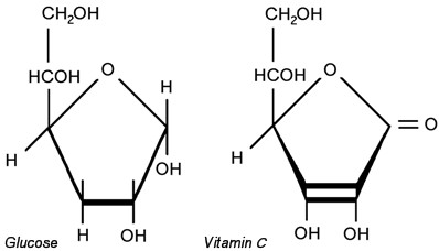
In the vast majority of animals, vitamin C is synthesized from glucose in only four metabolic steps. Hence, the molecular shape of vitamin C is remarkably similar to glucose. (Figure 1) Cancer cells will actively transport vitamin C into themselves, possibly because they mistake it for glucose. Another plausible explanation is that they are using the vitamin C as an antioxidant. Regardless, the vitamin C accumulates in cancer cells.
Figure 1
If large amounts of vitamin C are presented to cancer cells, large amounts will be absorbed. In these unusually large concentrations, the antioxidant vitamin C will start behaving as a pro-oxidant as it interacts with intracellular copper and iron. This chemical interaction produces small amounts of hydrogen peroxide.
Because cancer cells are relatively low in an intracellular anti-oxidant enzyme called catalase, the high dose vitamin C induction of peroxide will continue to build up until it eventually lyses the cancer cell from the inside out! This effectively makes high dose IVC a non-toxic chemotherapeutic agent that can be given in conjunction with conventional cancer treatments. Based on the work of several vitamin C pioneers before him, Dr. Riordan was able to prove that vitamin C was selectively toxic to cancer cells if given intravenously. This research was recently reproduced and published by Dr. Mark Levine at the National Institutes of Health.
As feared by many oncologists, small doses may actually help the cancer cells because small amounts of vitamin C may help the cancer cells arm themselves against the free-radical induced damage caused by chemotherapy and radiation. Only markedly higher doses of vitamin C will selectively build up as peroxide in the cancer cells to the point of acting in a manner similar to chemotherapy. These tumor-toxic dosages can only be obtained by intravenous administration.
Anticancer Res. 2009 Mar;29(3):809-15. High-dose vitamin C (ascorbic acid) therapy in the treatment of patients with advanced cancer.
Ohno S, Ohno Y, Suzuki N, Soma G, Inoue M.
Department of Complementary and Alternative Medicine Clinical R&D, Kanazawa University, Graduate School of Medical Science, Kanazawa, Ishikawa, 920-8640, Japan. satoshio@med.kanazawa-u.ac.jp
Vitamin C (ascorbic acid, ascorbate) has a controversial history in cancer treatment. Emerging evidence indicates that ascorbate in cancer treatment deserves re-examination. As research results concerning ascorbate pharmacokinetics and its mechanisms of action against tumor cells have been published, and as evidence from case studies has continued to mount that ascorbate therapy could be effective if the right protocols were used, interest among physicians and scientists has increased. In this review, high-dose vitamin C therapy in cancer treatment is re-evaluated.
1Department of Complementary and Alternative Medicine Clinical R&D, Kanazawa University, Graduate School of Medical Science, Ishikawa
2Department of Molecular Reproductive Biology, Kanazawa University, Graduate School of Medical Science, Ishikawa
3Institute for Health Sciences, Tokushima Bunri University, Tokushima
4Institute for Drug Delivery System, Tokyo University of Science, Chiba
5Department of Integrated and Holistic Immunology, Faculty of Medicine, Kagawa University, Kagawa, Japan
Correspondence to: Satoshi Ohno, Department of Complementary and Alternative Medicine Clinical R&D, Kanazawa University, Graduate School of Medical Science, 13-1 Takaramachi, Kanazawa, Ishikawa, 920-8640, Japan. Tel: +81 762652147, Fax: +81 762344247, e-mail: satoshio@med.kanazawa-u.ac.jp
Figure 1. Peak plasma vitamin C concentrations after oral or intravenous (IV) administration of vitamin C and cytotoxic effect of pharmacological vitamin C concentrations on tumor and normal cells. Plasma vitamin C concentrations that cause toxicity to cancer cells in vitro can be achieved clinically by intravenous, but not oral, administration of ascorbate.
Figure 2. Proposed mechanism of antitumor effects of vitamin C. The administration of more than 10 g of ascorbate is proposed to achieve plasma concentrations of 1 to 5 mM. At this time, vitamin C at high plasma concentration may function as a pro-oxidant. This occurs in the presence of free transition metals, such as copper and iron, which are reduced by ascorbate and, in turn, react with hydrogen peroxide (H2O2), leading to the formation of highly reactive and damaging hydroxyl radicals. As normal tissue receives adequate blood flow and is rich in antioxidant enzymes (e.g. catalase, glutathione peroxidase; GP) in the blood, any H2O2 formed will be immediately destroyed. Meanwhile, tumor tissue is often associated with reduced blood flow and antioxidant enzymes, and consequently formed H2O2 remains active leading to cell damage and death.
Footnotes
Received May 28, 2008.
Revision received July 31, 2008.
Accepted August 18, 2008.
Ann Oncol. 2008 Nov;19(11):1969-74. Epub 2008 Jun 9. Phase I clinical trial of i.v. ascorbic acid in advanced malignancy.
BACKGROUND: Ascorbic acid is a widely used and controversial alternative cancer treatment. In millimolar concentrations, it is selectively cytotoxic to many cancer cell lines and has in vivo anticancer activity when administered alone or together with other agents. We carried out a dose-finding phase I and pharmacokinetic study of i.v. ascorbic acid in patients with advanced malignancies. PATIENTS AND METHODS: Patients with advanced cancer or hematologic malignancy were assigned to sequential cohorts infused with 0.4, 0.6, 0.9 and 1.5 g ascorbic acid/kg body weight three times weekly. RESULTS: Adverse events and toxicity were minimal at all dose levels. No patient had an objective anticancer response. CONCLUSIONS: High-dose i.v. ascorbic acid was well tolerated but failed to demonstrate anticancer activity when administered to patients with previously treated advanced malignancies. The promise of this approach may lie in combination with cytotoxic or other redox-active molecules.
PMID: 18544557 [PubMed - indexed for MEDLINE]
THE CONTROVERSIAL STUDY THAT HIT THE NEWS in '08:
Cancer Res. 2008 Oct 1;68(19):8031-8. Vitamin C antagonizes the cytotoxic effects of antineoplastic drugs.
Vitamin C is an antioxidant vitamin that has been hypothesized to antagonize the effects of reactive oxygen species-generating antineoplastic drugs. The therapeutic efficacy of the widely used antineoplastic drugs doxorubicin, cisplatin, vincristine, methotrexate, and imatinib were compared in leukemia (K562) and lymphoma (RL) cell lines with and without pretreatment with dehydroascorbic acid, the commonly transported form of vitamin C. The effect of vitamin C on viability, clonogenicity, apoptosis, P-glycoprotein, reactive oxygen species (ROS), and mitochondrial membrane potential was determined. Pretreatment with vitamin C caused a dose-dependent attenuation of cytotoxicity, as measured by trypan blue exclusion and colony formation after treatment with all antineoplastic agents tested. Vitamin C given before doxorubicin treatment led to a substantial reduction of therapeutic efficacy in mice with RL cell-derived xenogeneic tumors. Vitamin C treatment led to a dose-dependent decrease in apoptosis in cells treated with the antineoplastic agents that was not due to up-regulation of P-glycoprotein or vitamin C retention modulated by antineoplastics. Vitamin C had only modest effects on intracellular ROS and a more general cytoprotective profile than N-acetylcysteine, suggesting a mechanism of action that is not mediated by ROS. All antineoplastic agents tested caused mitochondrial membrane depolarization that was inhibited by vitamin C. These findings indicate that vitamin C given before mechanistically dissimilar antineoplastic agents antagonizes therapeutic efficacy in a model of human hematopoietic cancers by preserving mitochondrial membrane potential. These results support the hypothesis that vitamin C supplementation during cancer treatment may detrimentally affect therapeutic response.
This report is available free online at www.liebertpub.com/act
In the Medical Journal Watch column of the latest issue, Jack Challem, a personal nutrition coach and nutrition author from Tucson, Arizona, and a regular contributor to the Journal, challenges the findings of a study published in Cancer Research (2008;68:8031-8038), in which the authors conclude that vitamin C given to mice or cultured cells treated with common anti-cancer drugs reduces the antitumor effects of the chemotherapeutic agents.
Challem points out two main problems with the study: the oxidized form of vitamin C (dehydroascorbic acid) and not actual vitamin C (ascorbic acid) was used; and in the mouse experiments, the animals were given toxic doses of dehydroascorbic acid, a compound that is not used as a dietary supplement in humans.
"This study and the subsequent headlines [it generated] were a grievous disservice to physicians and patients with cancer," says Challem. He adds that "considerable positive research…has shown striking benefits from high-dose vitamin C (ascorbic acid) in cancer cells and animals-and in actual human beings." High-dose intravenous vitamin C is a common form of alternative and complementary therapy for patients receiving chemotherapeutic drugs and is believed to help bring about tumor cell death. In addition, it may promote postsurgical healing by enhancing collagen formation, and increase tissue resistance to tumor spread.
Challem suggests that, "The ideal therapeutic approach would be to tailor individual treatment, including IV vitamin C, from a menu of options." http://www.liebertpub.com/act
P R Health Sci J. 2004 Jun;23(2):115-8. Intravenous vitamin C as a chemotherapy agent: a report on clinical cases.
Address correspondence to : Dr. Michael J. Gonzalez, University of Puerto Rico. Medical Sciences Campus, School of Public Health, Dept. Human Development,
Nutrition Program, RENAC 11 Project, PO BOX 365067, San Juan, and PR 00936 5067
Email: mgonzalez@rcm.upr.ed
Center for the Improvement of Human Functioning, RENAC Project, Wichita, KS, USA.
A series of seven cases are presented in which intravenous vitamin C has been used as antineoplastic agent in the treatment of different types of cancers. The cancers cases reviewed are the following: Renal cell carcinoma (2), Colorectal cancer (1), Pancreatic cancer (1), Non-Hodgkin's lymphoma (2) and breast cancer (1). Toxic reactions were not observed at these high doses of intravenous Vitamin C. All patients were prescreened for Glucose 6--phosphate dehydrogenase deficiency before administering intravenous Vitamin C in order to prevent hemolysis.
PMID: 15377059 [PubMed - indexed for MEDLINE]
Quote:
Intravenous vitamin c dose of more than 100 grams per day given in slow infusion are not toxic to cancer patients.
Moreover, some cancer patients have had complete remission of their cancer after a series of high dose intravenous vitamin C infusions. Also high doses of IV vitamin C have not interfered with the effect of
conventional therapy and may decrease toxicity of chemotherapy.
1. Riordan HD Jackson JA et al. Case study: high dose intravenous vitamin C in the treatment of a patient with adenocarcinoma of the kidney. J Orthomolec Med. 1990; 5:5-7.
2. Riordan HD Jackson JA et al. Case Study: High dose intravenous vitamin C in the treatment of a patient with renal cell carcinoma of the kidney. J Onhomolec Med 1998; 13:72-73.
3. Riordan NH Jackson JA et al. Case study: intravenous vitamin C in a terminal (breast cancer) cancer patient. J Orthomolec Med 1996;11:80-82.
J Transl Med. 2009 Aug 11;7:70. High dose concentration administration of ascorbic acid inhibits tumor growth in BALB/C mice implanted with sarcoma 180 cancer cells via the restriction of angiogenesis.
Yeom CH, Lee G, Park JH, Yu J, Park S, Yi SY, Lee HR, Hong YS, Yang J, Lee S.
Department of Palliative Medicine, Seoul St Mary's Hospital, The Catholic University of Korea, Seoul, 137-701, Korea. lymphych@hanmail.net
To test the carcinostatic effects of ascorbic acid, we challenged the mice of seven experimental groups with 1.7 x 10(-4) mol high dose concentration ascorbic acid after intraperitoneal administrating them with sarcoma S-180 cells. The survival rate was increased by 20% in the group that received high dose concentration ascorbic acid, compared to the control. The highest survival rate was observed in the group in which 1.7 x 10(-4) mol ascorbic acid had been continuously injected before and after the induction of cancer cells, rather than just after the induction of cancer cells. The expression of three angiogenesis-related genes was inhibited by 0.3 times in bFGF, 7 times in VEGF and 4 times in MMP2 of the groups with higher survival rates. Biopsy Results, gene expression studies, and wound healing analysis in vivo and in vitro suggested that the carcinostatic effect induced by high dose concentration ascorbic acid occurred through inhibition of angiogenesis.
CMAJ. 2006 Mar 28;174(7):937-42. Intravenously administered vitamin C as cancer therapy: three cases.
Padayatty SJ, Riordan HD, Hewitt SM, Katz A, Hoffer LJ, Levine M.
Molecular and Clinical Nutrition Section, Digestive Diseases Branch, National Institute of Diabetes and Digestive and Kidney Diseases, Bethesda, Md 20892-1372, USA.
Comment in:
Early clinical studies showed that high-dose vitamin C, given by intravenous and oral routes, may improve symptoms and prolong life in patients with terminal cancer. Double-blind placebo-controlled studies of oral vitamin C therapy showed no benefit. Recent evidence shows that oral administration of the maximum tolerated dose of vitamin C (18 g/d) produces peak plasma concentrations of only 220 micromol/L, whereas intravenous administration of the same dose produces plasma concentrations about 25-fold higher. Larger doses (50-100 g) given intravenously may result in plasma concentrations of about 14,000 micromol/L. At concentrations above 1000 micromol/L, vitamin C is toxic to some cancer cells but not to normal cells in vitro. We found 3 well-documented cases of advanced cancers, confirmed by histopathologic review, where patients had unexpectedly long survival times after receiving high-dose intravenous vitamin C therapy. We examined clinical details of each case in accordance with National Cancer Institute (NCI) Best Case Series guidelines. Tumour pathology was verified by pathologists at the NCI who were unaware of diagnosis or treatment. In light of recent clinical pharmacokinetic findings and in vitro evidence of anti-tumour mechanisms, these case reports indicate that the role of high-dose intravenous vitamin C therapy in cancer treatment should be reassessed.
PMID: 16567755 [PubMed - indexed for MEDLINE]
Pharmacologic doses of ascorbate act as a prooxidant and decrease growth of aggressive tumor xenografts in mice.
Chen Q, Espey MG, Sun AY, Pooput C, Kirk KL, Krishna MC, Khosh DB, Drisko J, Levine M.
Laboratory of Bioorganic Chemistry, National Institute of Diabetes and Digestive and Kidney Diseases, and Radiation Biology Branch, National Cancer Institute, National Institutes of Health, Bethesda, MD 20892, USA.
Comment in:
Ascorbic acid is an essential nutrient commonly regarded as an antioxidant. In this study, we showed that ascorbate at pharmacologic concentrations was a prooxidant, generating hydrogen-peroxide-dependent cytotoxicity toward a variety of cancer cells in vitro without adversely affecting normal cells. To test this action in vivo, normal oral tight control was bypassed by parenteral ascorbate administration. Real-time microdialysis sampling in mice bearing glioblastoma xenografts showed that a single pharmacologic dose of ascorbate produced sustained ascorbate radical and hydrogen peroxide formation selectively within interstitial fluids of tumors but not in blood. Moreover, a regimen of daily pharmacologic ascorbate treatment significantly decreased growth rates of ovarian (P < 0.005), pancreatic (P < 0.05), and glioblastoma (P < 0.001) tumors established in mice. Similar pharmacologic concentrations were readily achieved in humans given ascorbate intravenously. These data suggest that ascorbate as a prodrug may have benefits in cancers with poor prognosis and limited therapeutic options.
PMID: 18678913 [PubMed - indexed for MEDLINE]
Med Monatsschr Pharm. 2009 Jul;32(7):263-7. [Vitamin C in complementary oncology--update 2009]
[Article in German] Gröber U.
Akademie & Zentrum für Mikronährstoffmedizin, Zweigertstrasse 55, 45130 Essen. uwegroeber@gmx.net
The antioxidant perhaps most widely used in complementary oncology is vitamin C, particularly by intravenous injection. In light of the recent clinical pharmacokinetic findings, the in vitro evidence of anti-tumour mechanisms and some well-documented cases of advanced cancers the role of high-dose intravenous vitamin C therapy in cancer treatment should be reassessed. High dose intravenous vitamin C therapy may have benefits in patients with advanced cancers, and cancers with poor prognosis and limited therapeutic options, but further clinical studies regarding the safety and efficacy of this therapy are necessary, especially in Germany.
PMID: 19731754 [PubMed - indexed for MEDLINE]
Anticancer Res. 2009 Mar;29(3):809-15. High-dose vitamin C (ascorbic acid) therapy in the treatment of patients with advanced cancer.
Ohno S, Ohno Y, Suzuki N, Soma G, Inoue M.
Department of Complementary and Alternative Medicine Clinical R&D, Kanazawa University, Graduate School of Medical Science, Kanazawa, Ishikawa, 920-8640, Japan. satoshio@med.kanazawa-u.ac.jp
Vitamin C (ascorbic acid, ascorbate) has a controversial history in cancer treatment. Emerging evidence indicates that ascorbate in cancer treatment deserves re-examination. As research results concerning ascorbate pharmacokinetics and its mechanisms of action against tumor cells have been published, and as evidence from case studies has continued to mount that ascorbate therapy could be effective if the right protocols were used, interest among physicians and scientists has increased. In this review, high-dose vitamin C therapy in cancer treatment is re-evaluated.
PMID: 19414313 [PubMed - indexed for MEDLINE]
Proc Natl Acad Sci U S A. 2005 Sep 20;102(38):13604-9. Epub 2005 Sep 12. Pharmacologic ascorbic acid concentrations selectively kill cancer cells: action as a pro-drug to deliver hydrogen peroxide to tissues.
Chen Q, Espey MG, Krishna MC, Mitchell JB, Corpe CP, Buettner GR, Shacter E, Levine M.
Molecular and Clinical Nutrition Section, National Institute of Diabetes and Digestive and Kidney Diseases, National Institutes of Health, Bethesda, MD 20892, USA.
Human pharmacokinetics data indicate that i.v. ascorbic acid (ascorbate) in pharmacologic concentrations could have an unanticipated role in cancer treatment. Our goals here were to test whether ascorbate killed cancer cells selectively, and if so, to determine mechanisms, using clinically relevant conditions. Cell death in 10 cancer and 4 normal cell types was measured by using 1-h exposures. Normal cells were unaffected by 20 mM ascorbate, whereas 5 cancer lines had EC(50) values of <4 mM, a concentration easily achievable i.v. Human lymphoma cells were studied in detail because of their sensitivity to ascorbate (EC(50) of 0.5 mM) and suitability for addressing mechanisms. Extracellular but not intracellular ascorbate mediated cell death, which occurred by apoptosis and pyknosis/necrosis. Cell death was independent of metal chelators and absolutely dependent on H(2)O(2) formation. Cell death from H(2)O(2) added to cells was identical to that found when H(2)O(2) was generated by ascorbate treatment. H(2)O(2) generation was dependent on ascorbate concentration, incubation time, and the presence of 0.5-10% serum, and displayed a linear relationship with ascorbate radical formation. Although ascorbate addition to medium generated H(2)O(2), ascorbate addition to blood generated no detectable H(2)O(2) and only trace detectable ascorbate radical. Taken together, these data indicate that ascorbate at concentrations achieved only by i.v. administration may be a pro-drug for formation of H(2)O(2), and that blood can be a delivery system of the pro-drug to tissues. These findings give plausibility to i.v. ascorbic acid in cancer treatment, and have unexpected implications for treatment of infections where H(2)O(2) may be beneficial.
PMID: 16157892 [PubMed - indexed for MEDLINE]
Int J Oncol. 2009 Nov;35(5):1183-9. Fucoidan-Vitamin C complex suppresses tumor invasion through the basement membrane, with scarce injuries to normal or tumor cells, via decreases in oxidative stress and matrix metalloproteinases.
Saitoh Y, Nagai Y, Miwa N.
Cell-Death Control BioTechnology Laboratory, Faculty of Life and Environmental Sciences, Prefectural University of Hiroshima, Shobara, Hiroshima 727-0023, Japan.
Fucoidan (Fucdn) and vitamin C (VC) were saturatedly dissolved in water and lyophilized and thoroughly ethanol-rinsed until no detection for supernatant vitamin C to form the Fucdn-VC (1:0.23 wt/wt) inclusion body (Fucdn-VC-IB). Fucdn-VC-IB increased not only VC-stabilizing at 37 degrees C, but also hydroxyl-radical scavenging as shown by ESR/spin-trap method, more markedly than a mere mixture of Fucdn:VC (1:0.23 wt/wt). Invasion of human fibrosarcoma cells HT-1080 through the basement membrane was repressed by Fucdn-VC-IB of non-cytotoxic concentrations without significant inhibition to human skin dermal fibroblasts DUMS-16 cells. Fucdn-VC-IB suppressed the invasiveness-related gelatinases MMP-2/9, and diminished reactive oxygen species inside the cytoplasm around the nucleus, in HT-1080 cells as shown by electrophoretic zymography and the redox indicator NBT assay, respectively. Thus Fucdn-VC-IB markedly exhibits antioxidant and MMP-suppressing activities and preferentially inhibited tumor invasion without cytotoxicity to normal cells, and is suggested as a potent tumor-invasion suppressor.
PMID: 19787274 [PubMed - indexed for MEDLINE]
Int J Toxicol. 2009 Jan-Feb;28(1):33-42. Menadione reduction by pharmacological doses of ascorbate induces an oxidative stress that kills breast cancer cells.
Beck R, Verrax J, Dejeans N, Taper H, Calderon PB.
Université Catholique de Louvain, Louvain Drug Research Institute, Toxicology and Cancer Biology Research Group, PMNT Unit, Belgium. Oxidative stress generated by ascorbate-driven menadione redox cycling kills MCF7 cells by a concerted mechanism including glycolysis inhibition, loss of calcium homeostasis, DNA damage and changes in mitogen activated protein kinases (MAPK) activities. Cell death is mediated by necrosis rather than apoptosis or macroautophagy. Neither 3-methyladenine nor Z-VAD affects cytotoxicity by ascorbate/menadione (Asc/Men). BAPTA-AM, by restoring cellular capacity to reduce MTT, underlines the role of calcium in the necrotic process. Oxidative stress-mediated cell death is shown by the opposite effects of N-acetylcysteine and 3-aminotriazole. Moreover, oxidative stress induces DNA damage (protein poly-ADP-ribosylation and gamma-H2AX phosphorylation) and inhibits glycolysis. Asc/Men deactivates extracellular signal-regulated kinase (ERK) while activating p38, suggesting an additional mechanism to kill MCF7 cells. Since ascorbate is taken up by cancer cells and, due to their antioxidant enzyme deficiency, oxidative stress should affect cancer cells to a greater extent than normal cells. This differential sensitivity may have clinical applications.
PMID: 19482829 [PubMed - indexed for MEDLINE]
A Specific Combination Of Ascorbic Acid, Lysine, Proline And Epigallocatechin Gallate Inhibits Proliferation And Extracellular Matrix Invasion Of Various Human Cancer Cell Lines FULL TEXT
Shrirang P Netke, M Waheed Roomi, Vadim Ivanov, Aleksandra Niedzwiecki* and Matthias Rath
We found that a specific combination of ascorbic acid (AA, 100µM), proline (P, 140 µM) and lysine (L, 400 µM) had a significant antiproliferative and antimetastatic effect against some human cancer cell lines, breast (MDA-MB-231), colon (HCT116) and skin (melanoma, A2058). In the presence of this nutrient combination the invasion of the extracellular matrix by human breast cancer cells, melanoma cells and colon cancer cells was inhibited by 50%, 10% and 30% respectively. Addition of epigallocatechin gallate (EGCG) further enhanced this nutritional synergy producing a more pronounced inhibitory effect on both cellular proliferation and matrix invasion. The proliferation of breast cancer cells MDA-MB-231 in the presence of AA, P, L and 20µg/ml of EGCG was reduced to 74% and colon cancer cells HCT116 to 69% compared to the unsupplemented medium. The increase in concentration of EGCG to 50µg/ml did not cause much further reduction in the number of breast cancer cells. However it reduced proliferation of colon cancer cells to 4,6% and melanoma cells to 30% of the control. Matrigel invasion by breast cancer cells and human melanoma cells in the presence of AA, P, L and 20µg/ml of EGCG was stopped completely. At a similar concentration, invasion by colon cancer cells was reduced to 24,9%. However, the expression of matrix metalloproteinases (MMPs) –2 and –9 was not altered by this nutrient combination in melanoma cells as visualized by gelatinase zymography. MMP-2 and MMP-9 were significantly inhibited by EGCG in a dose dependent manner, and L, P, AA have no additional effect. Thus the combination of AA, P, L and EGCG shows great potential for control of cancer using a safe and multi-targeted approach.
Quote:
The antiproliferative activity of the nutrient combinations used in these studies varied with the type of cancer cells. In breast cancer cells, the combination of AA, P and L with EGCG had higher anti-proliferative effects than when these nutrients were used individually
Reverse endocrine therapy resistance?:
SABC 2009
67. Targeting Aldose Reductase: A Novel Strategy in Treating Endocrine Resistance Using Combination Therapy
Treating estrogen receptor-positive breast cancer tumors with a combination of fidarestat (an inhibitor of aldose reductase enzyme) and letrozole (an aromatase inhibitor) could delay or stop tumor resistance to endocrine therapy, according to data presented at the CTRC-AACR San Antonio Breast Cancer Symposium.
"Single agents are less effective," said Rajeshwar Rao Tekmal, Ph.D., professor of obstetrics and gynecology at the University of Texas Health Science Center at San Antonio. "Many tumors develop resistance, so this combination approach could prolong that window when endocrine therapy is effective."
About two-thirds of breast cancer tumors initially are hormone sensitive or estrogen receptor-positive and respond well to endocrine therapy. However, close to half of those tumors develop resistance to endocrine therapy, said Tekmal.
In this preclinical study, researchers treated estrogen receptor-positive tumors already resistant to letrozole with letrozole and fidarestat. As an inhibitor of aldose reductase enzyme, fidarestat blocks the metabolism of glucose in cancer cells. Together, the combination effectively re-sensitized the cells to letrozole, allowing for effective endocrine therapy and more cell death.
Researchers believe increased glucose metabolism (polyol accumulation) contributes to oxidative stress, which, in turn, could alter intracellular signalling by affecting the regulation of protein kinases that are known to be involved in therapy resistance. Blocking the path of glucose metabolism may help to restore sensitivity to endocrine therapies or it may stop or delay the development resistance endocrine therapies in first place.
While this is a preclinical study, Tekmal believes it could lead to future drug treatments that will make endocrine therapy more effective for longer periods of time.
"This is a very promising study showing that combination treatments seem to work on resistance and re-sensitizing tumors that are resistant to endocrine therapies," he said.
Listen to a recording of the teleconference:
Download* the mp3 of the teleconference (13.2 MB, 58 minutes and 04 seconds)
Vitamin C: an aldose reductase inhibitor that normalizes erythrocyte sorbitol in insulin-dependent diabetes mellitus
J. J. Cunningham, P. L. Mearkle and R. G. Brown
Department of Nutrition, University of Massachusetts, Amherst 01003-1420.
OBJECTIVE: Diabetic hyperglycemia promotes sorbitol production from glucose via aldose reductase. Since the intracellular accumulation of sorbitol, or its sequelae, are postulated to contribute to the progression of chronic diabetic complications, aldose reductase inhibitors (ARI) offer therapeutic promise. Others have shown that vitamin C at pharmacologic doses decreases erythrocyte (RBC) sorbitol. We examined whether smaller, physiologic doses of vitamin C were also effective in individuals with insulin-dependent diabetes mellitus (IDDM) and whether vitamin C was an ARI in vitro. METHODS: Vitamin C supplements (100 or 600 mg) were taken daily for 58 days by young adults with IDDM and nondiabetic adults in an otherwise free-living design. Diabetic control was monitored by fasting plasma glucose, glycosylated hemoglobin, and glycosuria and was moderate to poor throughout the study. RBC sorbitol was measured at baseline and again at 30 and 58 days. Three-day dietary records and 24-hour urine collections were performed for each sampling day. RESULTS: RBC sorbitol levels were significantly elevated in IDDMs, on average doubled, despite their more than adequate dietary intakes of vitamin C and normal plasma concentrations. Vitamin C supplementation at either dose normalized the RBC sorbitol in IDDMs within 30 days. This correction of sorbitol accumulation was independent of changes in diabetic control. Furthermore, our in vitro studies show that ascorbic acid inhibited aldose reductase activity. CONCLUSIONS: Vitamin C supplementation is effective in reducing sorbitol accumulation in the erythrocytes of diabetics. Given its tissue distribution and low toxicity, we suggest a superiority for vitamin C over pharmaceutic ARIs.
Cancer Chemother Pharmacol. 2010 Mar 6. [Epub ahead of print] The antioxidant ascorbic acid mobilizes nuclear copper leading to a prooxidant breakage of cellular DNA: implications for chemotherapeutic action against cancer.
Ullah MF, Khan HY, Zubair H, Shamim U, Hadi SM.
Department of Biochemistry, Faculty of Life Sciences, AMU, Aligarh, UP, 202002, India.
PURPOSE: Ascorbic acid is an essential micronutrient and is considered to have an antioxidant function in living systems. For the past several decades, ascorbic acid has been the subject of considerable interest as an anticancer agent. Several studies have shown that ascorbic acid is cytotoxic to a variety of cancer cells, whereas normal cells are relatively resistant to such cytotoxic action. In this study, we propose a putative molecular mechanism that accounts for the preferential cytotoxicity of ascorbic acid against cancer cells. METHODS: Standard and lysed version of alkaline single-cell gel electrophoresis (Comet assay); ferrous oxidation-xylenol orange (FOX) assay. RESULTS: We show that ascorbic acid acts as a prooxidant and leads to oxidative DNA breakage in lymphocytes and lymphocyte nuclei. Scavengers of reactive oxygen species were able to inhibit ascorbic acid-induced DNA breakage, suggesting the involvement of reactive oxygen species in this reaction. We further show that such DNA breakage is inhibited by both iron and copper chelators in cells, whereas in nuclei, similar inhibition was achieved only by copper chelators, indicating an important role of chromatin-bound copper in the prooxidant cellular DNA breakage by ascorbic acid. CONCLUSION: We propose that the copper-dependent cellular redox status is an important element in the cytotoxic action of ascorbic acid against cancer cells. It is well established that cellular copper levels are considerably elevated in various malignancies. Therefore, cancer cells may be more subject to electron transfer between copper and ascorbate to generate reactive oxygen species. In light of these observations and those in literature, in this paper we explain that the preferential cytotoxicity of ascorbic acid against cancer cells is the result of elevated copper levels in such cells. Further, this study identifies nuclear copper as a novel molecular target for cytotoxic action of ascorbic acid, which has implications for its chemotherapeutic properties against cancer.
PMID: 20213077 [PubMed - as supplied by publisher]
Interesting how an argument/research can be skewed so easily, isn't it? Why not use the form most readily available to the public and in doses commonly used by them? Doesn't make sense to me. Am I missing something here, Rich?
__________________
Smile On!
Laurel
Dx'd w/multifocal DCIS/IDS 3/08
7mm invasive component
Partial mast. 5/08
Stage 1b, ER 80%, PR 90%, HER-2 6.9 on FISH
0/5 nodes
4 AC, 4 TH finished 9/08
Herceptin every 3 weeks. Finished 7/09
Tamoxifen 10/08. Switched to Femara 8/09
Bilat SPM w/reconstruction 10/08
Clinical Trial w/Clondronate 12/08
Stopped Clondronate--too hard on my gizzard!
Switched back to Tamoxifen due to tendon pain from Femara
15 Years NED
I think I just might hang around awhile....
(I assume you referring to supplementation not monotherapy)
Dunno..
Not the first time I've seen research inexplicably determined to be irrelevant and/or undermining. Same thing happened with Metformin showing problems in mice at 40 times the usual serum level..in mice. Good for simulating overdose or someone with kidney shutdown. Not too elucidating. I don't like conspiracy theories but I'm not too sure incompetence is more reassuring. But..I'm sure I don't get the nuances of it all
Seems like they had some good data with good old ascorbic acid.
__________________
Smile On!
Laurel
Dx'd w/multifocal DCIS/IDS 3/08
7mm invasive component
Partial mast. 5/08
Stage 1b, ER 80%, PR 90%, HER-2 6.9 on FISH
0/5 nodes
4 AC, 4 TH finished 9/08
Herceptin every 3 weeks. Finished 7/09
Tamoxifen 10/08. Switched to Femara 8/09
Bilat SPM w/reconstruction 10/08
Clinical Trial w/Clondronate 12/08
Stopped Clondronate--too hard on my gizzard!
Switched back to Tamoxifen due to tendon pain from Femara
15 Years NED
I think I just might hang around awhile....
Updated above with phase I IV ascorbic acid monotherapy in advanced Ca: no responses. May be of use with other therapies or in earlier cancer.
Anticancer Res. 2003 Jul-Aug;23(4):3279-87.
The in vitro antitumor activity of vitamins C and K3 against ovarian carcinoma.
von Gruenigen VE, Jamison JM, Gilloteaux J, Lorimer HE, Summers M, Pollard RR, Gwin CA, Summers JL.
Department of Obstetrics and Gynecology, SUMMA Health System, 525 E. Market Street, Akron, OH 44309, USA. vivian.vongruenigen@uhhs.com
BACKGROUND: The objective was to evaluate the cytotoxic effect and mechanism of action of vitamins C (VC) and K3 (VK3) on ovarian carcinoma. MATERIALS AND METHODS: Cytotoxicity assays were performed on ovarian cancer cell lines with VC, VK3 or a VC/VK3 combination. FIC index was employed to evaluate synergism. Flow cytometry was accomplished at 90% cytotoxic doses. Light, transmission electron microscopy and DNA isolation were performed. RESULTS: Antitumor activity was exhibited by both VC, VK3 and VC/VK3. VC/VK3 demonstrated synergistic activity. VC/VK3 may induce a G1 block in the cell cycle. Combined vitamin treatment resulted in cells that maintain apparently intact nuclei while extruding pieces of organelle-free cytoplasm. Degradation of chromosomal DNA was observed. CONCLUSION: Cell death (autoschizis) displayed characteristics of both apoptosis and necrosis. The cytotoxic effects observed may enable vitamins C and K3 to play an adjuvant role in the treatment of ovarian cancer.
PMID: 12926064 [PubMed - indexed for MEDLINE]
Eur J Med Chem. 2003 May;38(5):451-7.
The association of vitamins C and K3 kills cancer cells mainly by autoschizis, a novel form of cell death. Basis for their potential use as coadjuvants in anticancer therapy.
Deficiency of alkaline and acid DNase is a hallmark in all non-necrotic cancer cells in animals and humans. These enzymes are reactivated at early stages of cancer cell death by vitamin C (acid DNase) and vitamin K(3) (alkaline DNase). Moreover, the coadministration of these vitamins (in a ratio of 100:1, for C and K(3), respectively) produced selective cancer cell death. Detailed morphological studies indicated that cell death is produced mainly by autoschizis, a new type of cancer cell death. Several mechanisms are involved in such a cell death induced by CK(3), they included: formation of H(2)O(2) during vitamins redox cycling, oxidative stress, DNA fragmentation, no caspase-3 activation, and cell membrane injury with progressive loss of organelle-free cytoplasm. Changes in the phosphorylation level of some critical proteins leading to inactivation of NF-kappaB appear as main intracellular signal transduction pathways. The increase knowledge in the mechanisms underlying cancer cells death by CK(3) may ameliorate the techniques of their in vivo administration. The aim is to prepare the introduction of the association of vitamins C and K(3) into human clinics as a new, non-toxic adjuvant cancer therapy.
PMID: 12767595 [PubMed - indexed for MEDLINE]
Br J Cancer. 2010 Mar 23. [Epub ahead of print]
alpha-Tocopheryl succinate promotes selective cell death induced by vitamin K3 in combination with ascorbate.
Tomasetti M, Strafella E, Staffolani S, Santarelli L, Neuzil J, Guerrieri R.
Department of Molecular Pathology and Innovative Therapies, Polytechnic University of Marche, Ancona 60020, Italy.
Background: A strategy to reduce the secondary effects of anti-cancer agents is to potentiate the therapeutic effect by their combination. A combination of vitamin K3 (VK3) and ascorbic acid (AA) exhibited an anti-cancer synergistic effect, associated with extracellular production of H(2)O(2) that promoted cell death. Methods: The redox-silent vitamin E analogue alpha-tocopheryl succinate (alpha-TOS) was used in combination with VK3 and AA to evaluate their effect on prostate cancer cells.
Results: Prostate cancer cells were sensitive to alpha-TOS and VK3 treatment, but resistant to AA upto 3.2 mM. When combined, a synergistic effect was found for VK3-AA, whereas alpha-TOS-VK3 and alpha-TOS-AA combination showed an antagonist and additive effect, respectively. However, sub-lethal doses of AA-VK3 combination combined with a sub-toxic dose of alpha-TOS showed to induce efficient cell death that resembles autoschizis. Associated with this cell demise, lipid peroxidation, DNA damage, cytoskeleton alteration, lysosomal-mitochondrial perturbation, and release of cytochrome c without caspase activation were observed. Inhibition of lysosomal proteases did not attenuate cell death induced by the combined agents. Furthermore, cell deaths by apoptosis and autoschizis were detected.
Conclusion:These finding support the emerging idea that synergistic combinations of some agents can overcome toxicity and other side-effects associated with high doses of single drugs creating the opportunity for therapeutically relevant selectivity.
British Journal of Cancer advance online publication, 23 March 2010; doi:10.1038/sj.bjc.6605617 www.bjcancer.com.
PMID: 20332775 [PubMed - as supplied by publisher]
Dis Esophagus. 2009 Nov 23. [Epub ahead of print]
Pilot translational study of dietary vitamin C supplementation in Barrett's esophagus.
The transcription factor Nuclear factor kappa B (NF-kappaB) is central to the regulation of genes encoding for mediators of inflammation and carcinogenesis. In the esophagus, NF-kappaB is progressively activated from inflammation to Barrett's metaplasia and adenocarcinoma. Vitamin C, an antioxidant, can inhibit NF-kappaB in in vitro models, and the aim of this study was to prospectively assess the effect of supplemental vitamin C on NF-kappaB and associated cytokines in patients with Barrett's esophagus. Twenty-five patients with long-segment Barrett's and specialized intestinal metaplasia received dietary vitamin C (1000 mg/day) orally for four weeks, and had pre- and post-vitamin C endoscopic biopsies. NF-kappaB activity (activated p50 and p65 subunits) of nuclear extracts was assessed using the Active Motif NF-kappaB assay, and cytokines and growth factors were measured using the Evidence Investigator biochip array. NF-kappaB and related pro-inflammatory cytokines and growth factors (IL-8, VEGF, IL-10) were activated in all Barrett's tissue pre-treatment. Down-regulation in activated NF-kappaB and cytokines was observed in 8/25 (35%) patients. Dietary vitamin C supplementation may down-regulate pro-inflammatory markers in a subset of Barrett's patients. Further studies with larger numbers of endpoints will be needed to further evaluate this effect.
PMID: 19930402 [PubMed - as supplied by publisher]
By Martin Johnston
4:00 AM Tuesday Jul 20, 2010 New Zealand researchers have established that vitamin C can help to block the growth of cancer cells - an important experimental finding they expect could be quickly adopted into cancer treatment.
The role of vitamin C in cancer treatment has been controversial for decades, with contradictory findings from various studies. In an international review of 20 human trials of vitamin C and other "anti-oxidant" supplements, the influential Cochrane Collaboration found no convincing evidence that they could prevent gastro-intestinal cancers - and said they "even seem to increase mortality".
But now a team from Otago University at Christchurch, in a paper published in leading international journal Cancer Research, have shown that vitamin C has a role in controlling tumour growth.
They say their study of tumorous and normal tissue samples from women with cancer of the uterine lining provides the first direct evidence of a link between vitamin C and a protein called HIF-1.
HIF (hypoxia inducible factor)-1 is considered a key protein in tumour survival. High activity of it promotes tumour growth and resistance to chemotherapy and radiotherapy and is linked with a poor prognosis for patients.
The Christchurch study, led by Associate Professor Margreet Vissers, of the university's Free Radical Research Group, found that high-grade tumours had around 40 per cent less vitamin C than matched, adjacent, normal tissue.
The researchers say their study suggests that restoring the vitamin C levels in tumours would limit factors that promote tumour growth, and recommend animal trials to test the hypothesis.
Professor Vissers said the study suggested it would be beneficial for people with cancer cells to have more vitamin C. It could help restrict the rate of tumour growth, increase responsiveness to chemotherapy and might prevent formation of solid tumours.
"There's enough information now for people to be seriously thinking about doing this, to apply this to the clinic or be setting up some clinical trials," she told the Herald yesterday.
"Anti-oxidant supplementation may not end up delivering any more vitamin C to the tumour.
"Just supplementing people may not actually have the effect that you want because you haven't done it in the right way," Professor Vissers said.
She said vitamin C levels in the body could be raised only to a certain level by oral supplementation.
Intravenous injections could achieve a higher level.
"That's the question: what's the best way to deliver vitamin C to the tumour."
By Martin Johnston
It is suspected that vitamin C is good for normal tissue because it protects the mitochondria and extends cell life. However, that's not what you want in cancer cells, where you want cell death to be speeded up so the tumor shrinks. In other words, vitamin C appears to protect the mitochondria from extensive damage, thus saving the cell. All anticancer drugs work, directly or indirectly, to disrupt the mitochondria to push cell death (Cancer Res 2008;68(19):8031–8).
However, vitamin C can impede the growth of some types of tumors, although not in the way some scientists had suspected. New research, published in the journal Cancer Cell, supported the general notion that vitamin C and other antioxidants can slow tumor growth, but at a mechanism different from the one many experts had suspected (undermining a tumor's ability to grow under certain conditions).
Earlier studies were conducted with vitamin C "supplements" administered orally and did not demonstrate a clinical benefit to cancer patients. Newer studies, are being conducted with "intravenous" vitamin C as a cancer treatment and may lend credence to alternate cancer care.
New research with mice suggests that intravenous doses of vitamin C could reduce the size of cancerous tumors in people. It would likely be used in combination with other drugs. In other words, vitamin C will be used as a drug, not as a nutrient. The findings must be confirmed in humans.
The first phase of trials are being performed to determine the optimal dose for the patients and to learn whether the treatment is safe and can be tolerated. Additional studies over several years will be needed to demonstrate whether it is effective.
It's ironic, clinical trials are proceeding only with patients who have exhausted all other conventional treatment options, instead of enrolling anyone who would want to enroll.


 Linear Mode

